Modified Biopsy Technique for Squamous Cell Carcinoma on the Forehead: A Detailed Approach
Squamous cell carcinoma (SCC) is a common type of skin cancer that arises from the keratinocytes of the epidermis. While it can occur on any part of the body, SCC on the forehead presents unique challenges due to the region’s high visibility, thin skin, and proximity to critical structures such as the eyes and facial nerves. Accurate diagnosis and treatment planning are essential to ensure optimal outcomes, and biopsy remains the gold standard for confirming SCC. However, traditional biopsy techniques may not always be suitable for lesions on the forehead, particularly when cosmetic outcomes and tissue preservation are priorities. This article explores a modified biopsy technique tailored for SCC on the forehead, emphasizing precision, minimal invasiveness, and aesthetic considerations.
Challenges of Forehead Biopsies
The forehead is a cosmetically sensitive area, and any surgical intervention must balance diagnostic accuracy with the preservation of form and function. Traditional biopsy methods, such as punch or shave biopsies, can sometimes result in scarring, tissue distortion, or incomplete sampling, especially in cases where the lesion is large or ill-defined. Additionally, the forehead’s thin skin and underlying bone structure require careful handling to avoid complications such as bleeding, infection, or damage to adjacent structures.
The Modified Biopsy Technique
The modified biopsy technique for SCC on the forehead is designed to address these challenges by incorporating principles of precision, minimal tissue disruption, and aesthetic preservation. The approach involves the following steps:
1. Pre-Biopsy Assessment
- Clinical Evaluation: A thorough clinical examination is performed to assess the size, depth, and borders of the lesion. Dermoscopy may be used to evaluate the lesion’s characteristics and guide the biopsy site selection.
- Imaging: In cases where deep invasion is suspected, imaging modalities such as ultrasound or MRI may be employed to assess the extent of the tumor and its relationship to underlying structures.
2. Anesthesia and Preparation
- Local Anesthesia: The area is anesthetized using a local anesthetic, such as lidocaine with epinephrine, to minimize bleeding and discomfort.
- Sterile Technique: The biopsy site is cleaned and draped to maintain a sterile field, reducing the risk of infection.
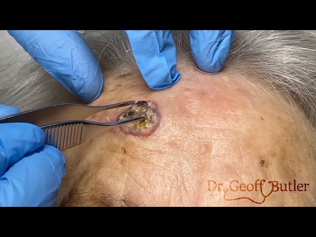
3. Incisional Biopsy with Margin Mapping
- Precise Incision: A scalpel is used to make a small, full-thickness incision within the lesion, ensuring that the sample includes both the central and peripheral portions of the tumor. This approach allows for accurate histopathological assessment of the tumor’s margins and depth.
- Margin Mapping: To ensure complete sampling, multiple small incisions may be made around the lesion’s periphery. This technique, known as margin mapping, helps identify any subclinical extension of the tumor and guides subsequent treatment planning.
4. Tissue Handling and Closure
- Gentle Handling: The biopsy specimen is carefully handled to avoid crush artifacts, which can compromise histopathological analysis.
- Layered Closure: The wound is closed in layers using fine, non-absorbable sutures to minimize tension and reduce the risk of scarring. In some cases, absorbable sutures may be used for the deeper layers to provide additional support.
5. Post-Biopsy Care
- Wound Care: Patients are instructed on proper wound care, including keeping the area clean and applying topical antibiotics to prevent infection.
- Follow-Up: A follow-up appointment is scheduled to review the biopsy results and discuss further treatment options, such as surgical excision, radiation therapy, or Mohs micrographic surgery.
Advantages of the Modified Technique
- Enhanced Diagnostic Accuracy: By incorporating margin mapping, the modified technique ensures comprehensive sampling of the lesion, reducing the risk of missed or incomplete diagnoses.
- Aesthetic Preservation: The use of precise incisions and layered closure minimizes scarring and preserves the natural contours of the forehead.
- Minimized Complications: The technique’s emphasis on gentle tissue handling and sterile technique reduces the risk of bleeding, infection, and damage to adjacent structures.
Conclusion
The modified biopsy technique for squamous cell carcinoma on the forehead represents a refined approach that balances diagnostic accuracy with aesthetic and functional considerations. By incorporating principles of precision, minimal invasiveness, and careful tissue handling, this technique offers a reliable method for diagnosing SCC while preserving the patient’s quality of life. As with any medical procedure, the technique should be tailored to the individual patient’s needs and performed by a skilled dermatologist or surgeon to ensure optimal outcomes.